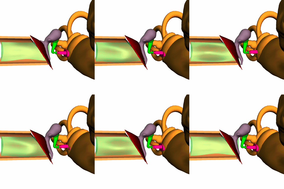
New research at Bristol takes a closer look and separates out the parts more clearly. So I’ll do the same.
First we see Katy walking along a branch, and pausing when she hears something interesting…

What does she hear?

Perhaps something in the air, or perhaps a vibration on the stem that indicates a tasty insect at work or a matable male singing Our Song.

Where are the ears?

On the second joint of each front leg. Each leg has two eardrums facing roughly 90 degrees apart.

Here’s a transverse view of the joint in the same position. The Crista is the framework that holds the sensory hair cells, and merges their signals into a cable leading to the brain. The trachea is actually divided into two parallel pipes, and each tympanum pushes on one of the pipes. The trachea ultimately runs into one of the spiracles (lungs) on this side of the abdomen. Most likely the trachea serves the same purpose as our Eustachian tube, equalizing major changes of pressure so the tympanum can move more freely. Some researchers think the trachea is the main inlet for sound, but this seems unlikely.

How do the two tympani respond to waves in the air? Here we have a major difference. Our eardrums are at the end of a canal, so sound from all directions is narrowed down into a single inward-moving wave.

Katy’s tympani are fully exposed, with their length parallel to the row of sensory haircells. So the sensing organ can read the direction of sound.

The row of 50 sensory cells is tuned just as ours is tuned. The cilia at one end are shorter, and the cells are smaller, so they will respond to higher frequencies.
Most of the tuning in our cochlea is done by resonance within the fluid. A mix of frequencies is coming in via the tympanum, and the various harmonics and formants bounce through the chambers differently.
Some frequencies form a traveling wave, which runs through the chambers with no net force at one point:

And some frequencies form a standing wave at one location, giving strong motion to one group of hair cells:

Here’s a cross section of our cochlea while it receives a standing wave. We also have two parallel tubes, but they aren’t sensing two sides of the universe; they’re just carrying different types of fluid. Unlike the katydid, we have two separate groups of haircells. The inner cells (left here) are the actual sensors. The outer cells (right) are effectively muscles, exerting feedback counterforce on the upper membrane to damp down steady or uninteresting sounds.


